

Wisdom Teeth Removal Chino: Procedure and Recovery
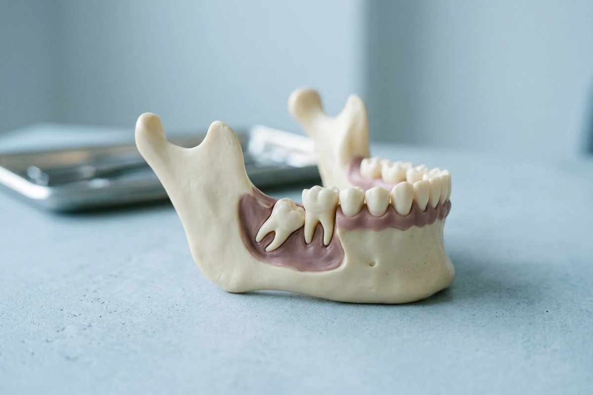
Evaluating third molars helps protect surrounding teeth and oral health. Discover how wisdom teeth removal Chino clinicians provide addresses impacted teeth, procedural steps, sedation choices, and practical recovery guidelines for lasting comfort.
Understanding Third Molars and Extraction Needs
Third molars represent the last set of adult teeth to develop in the dental arch. When jaw anatomy prevents proper positioning, surgical extraction becomes necessary to prevent localized oral complications, maintain bite alignment, and protect adjacent tooth structures from progressive oral health damage.[1][2]
The Mechanics of Dental Impaction
An impacted wisdom tooth dentist evaluates cases where third molars lack adequate space to erupt naturally through the alveolar bone and gingival tissue. Impaction happens when jaw dimensions restrict normal emergence or teeth develop along an abnormal trajectory, causing them to remain trapped beneath the gums or press into adjacent teeth.[1][2]
Clinical Classifications of Impacted Teeth
Clinicians classify impactions by tissue obstruction and developmental angulation. Soft-tissue impactions occur when crowns clear bone but remain covered by gingiva, whereas partial or complete bony impactions stay enclosed within alveolar bone. Angulations range from horizontal or vertical alignments to mesioangular tilts toward neighboring teeth or distoangular angles pointing backward.[2][3]
Factors Influencing Surgical Timing and Candidacy
Determining candidacy for surgical intervention depends on developmental maturity, anatomical positioning, and existing structural pathology. Evaluating these critical clinical elements ensures that treatment decisions minimize surgical invasiveness while promoting predictable, efficient healing outcomes across various stages of patient development.[1][3]
Developmental Timing and Patient Age
Evaluating third molars during late adolescence or early adulthood provides distinct clinical advantages. During this developmental window between ages seventeen and twenty-five, tooth roots are typically only two-thirds formed and adjacent alveolar bone remains relatively pliable, which lowers procedural complexity and significantly shortens post-operative tissue recovery timelines.[1][3]
Pathological Risks to Adjacent Structures
Retaining impacted third molars introduces clinical risks to neighboring dentition. Trapped molars can foster destructive periodontal bone loss, cause external root resorption on adjacent second molars, or promote the development of follicular pathologies such as dentigerous cysts, which expand silently and erode surrounding supportive jawbone structures over extended periods.[1][3]
Recognizing Common Symptoms and Clinical Warnings
Symptoms associated with impacted molars range from subtle, periodic pressure to acute bacterial infections. Recognizing localized signs allows patients to address developing issues before deep structural bone damage or widespread soft-tissue infections compromise general oral function and comfort.[1][3]
Characteristic Indicators of Third Molar Distress
Partially erupted molars frequently trap bacteria and food debris under overlying gingival tissue, causing pericoronitis, localized swelling, tender gums, and persistent halitosis. Patients may also experience dull posterior jaw pain or trismus, which is difficulty opening the mouth, though some problematic impactions remain entirely asymptomatic until advanced damage occurs.[1][3]
Comprehensive Clinical Evaluation and Diagnostic Imaging
A thorough clinical evaluation establishes the exact anatomical relationship between third molars and vital oral structures. Dental professionals employ visual assessments alongside targeted radiographic diagnostic tools to chart extraction pathways, assess nerve proximity, and design individualized, safe surgical plans.[1][2][3]
Diagnostic Imaging and Anatomical Mapping
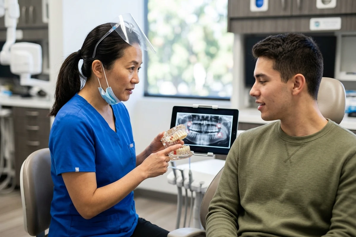
A panoramic radiograph serves as the primary diagnostic tool to evaluate root anatomy, tooth angulation, and proximity to the maxillary sinus or inferior alveolar nerve. If roots closely approximate sensitive neural pathways, cone-beam computed tomography provides precise three-dimensional views, helping clinicians mitigate risks prior to performing wisdom tooth extraction Chino procedures.[1][2][3]
Treatment Approaches and Surgical Alternatives
Management of third molars extends beyond standard extraction to include non-surgical observation and conservative surgical adaptations. Selecting the appropriate intervention requires balancing active disease treatment against potential neurological risks, surgical invasiveness, and the patient's comprehensive long-term dental needs.[1][3]
Active Surveillance Protocols
Active surveillance represents a conservative alternative for completely asymptomatic, disease-free wisdom teeth embedded deep within alveolar bone. When surgical removal introduces substantial risks, such as potential nerve injury, clinicians may recommend annual visual exams and routine radiographic imaging to monitor the area continuously without undergoing preemptive surgical extraction.[1][3]
Coronectomy for Nerve Protection
When diagnostic scans show third molar roots intimately entwined with the inferior alveolar nerve canal, an oral surgeon Chino patients consult may perform a coronectomy. This partial odontectomy removes only the enamel crown while leaving healthy, stable root tips undisturbed in bone, substantially decreasing the likelihood of nerve paresthesia.[2][3]
Surgical Extraction Procedure and Sedation Options
The extraction procedure follows established surgical techniques designed to remove impacted teeth efficiently while preserving surrounding bone and soft tissue. Careful anesthesia administration, strategic sectioning methods, and surgical debridement combine to produce predictable, comfortable clinical outcomes during every phase.[1][2][3]
Anesthesia and Comfort Management
Surgical teams tailor anesthesia approaches to clinical complexity and individual patient health profiles. Local anesthesia numbs the treatment site entirely, while nitrous oxide, intravenous sedation, or general anesthesia can be administered to provide deep relaxation. These tailored options maintain patient comfort throughout complex bone removal or multi-tooth procedures.[1][3]
Surgical Steps for Impacted Tooth Removal
The surgical sequence follows systematic, conservative techniques designed to remove impacted third molars efficiently while safeguarding adjacent structures. After administering anesthesia, clinicians carefully access the operative site, relieve anatomical resistance, and close the wound to establish favorable conditions for post-operative tissue healing and reliable blood clot formation.[1][2][3]
- Incising and reflecting overlying gingival tissue to expose the alveolar bone and embedded tooth crown.
- Removing thin layers of overlying bone and gently sectioning the tooth into smaller segments for conservative delivery.
- Elevating the tooth fragments carefully, thoroughly irrigating the surgical socket, and placing sutures to secure the tissue.
Post-Operative Recovery and Healing Guidelines
Post-operative recovery focuses on stabilizing the surgical site, managing normal tissue inflammation, and protecting the primary blood clot. Following established aftercare recommendations ensures a smooth transition toward normal dietary habits, consistent oral hygiene routines, and uneventful soft-tissue healing.[1][3]
Protecting the Surgical Site and Blood Clot
Preserving the initial blood clot within the extraction socket is vital for preventing alveolar osteitis, commonly known as dry socket. Patients must avoid vigorous mouth rinsing, forceful spitting, drinking through straws, and tobacco smoking during early healing, as mechanical suction can dislodge the protective clot and expose sensitive bone.[1][3]
Managing Swelling and Resuming Oral Hygiene
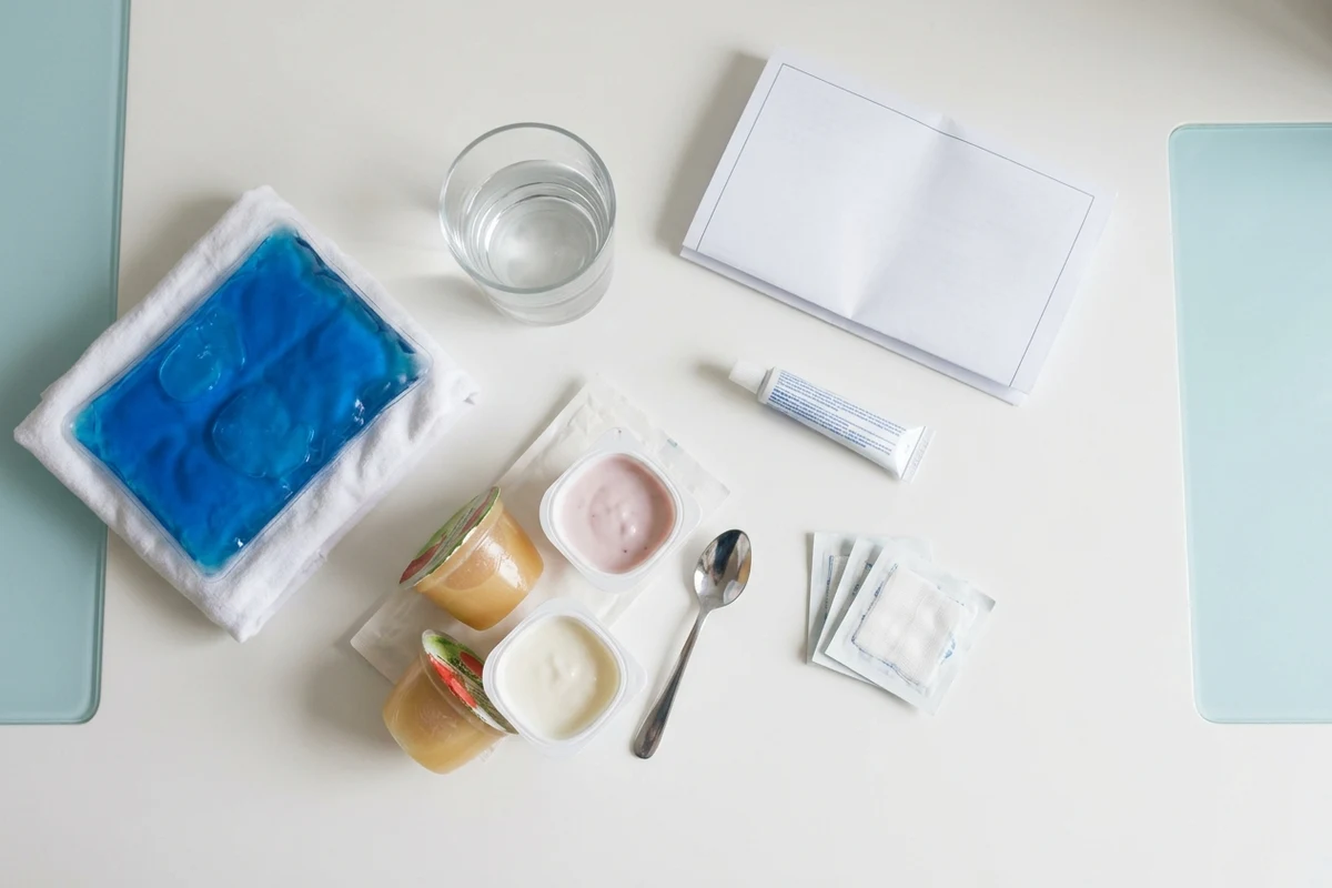
Post-operative facial swelling and mild jaw stiffness typically reach peak intensity between forty-eight and seventy-two hours after surgery before gradually resolving over seven to ten days. Patients should maintain a soft-food diet and resume gentle oral hygiene, incorporating warm saline rinses twenty-four hours post-surgery to maintain cleanliness.[1][3]
Managing Complications and Arranging Professional Care
Although wisdom tooth removal is routinely performed with high success rates, understanding potential complications empowers patients to recognize issues early. Timely communication with your clinical team ensures minor healing disruptions are addressed promptly before developing into more serious complications.[1][3]
Recognizing Post-Extraction Complications
Contact the clinic if throbbing discomfort intensifies three to five days post-surgery, which signals dry socket, or if persistent drainage, foul odor, or localized numbness develops. Severe swelling, persistent fever, or uncontrolled bleeding warrant emergency wisdom tooth removal evaluations to treat spreading infection and safeguard oral health.[1][3]
Sources
- mouthhealthy.org — American Dental Association
- ada.org — American Dental Association
- nih.gov — pmc.ncbi.nlm.nih.gov
Common questions
Frequently asked questions
Talk with a dentist about your next step
Contact One Ounce Dental today to schedule an evaluation and discuss your wisdom teeth removal questions with our caring dental team. We are here to support your oral health needs.
More Articles
View all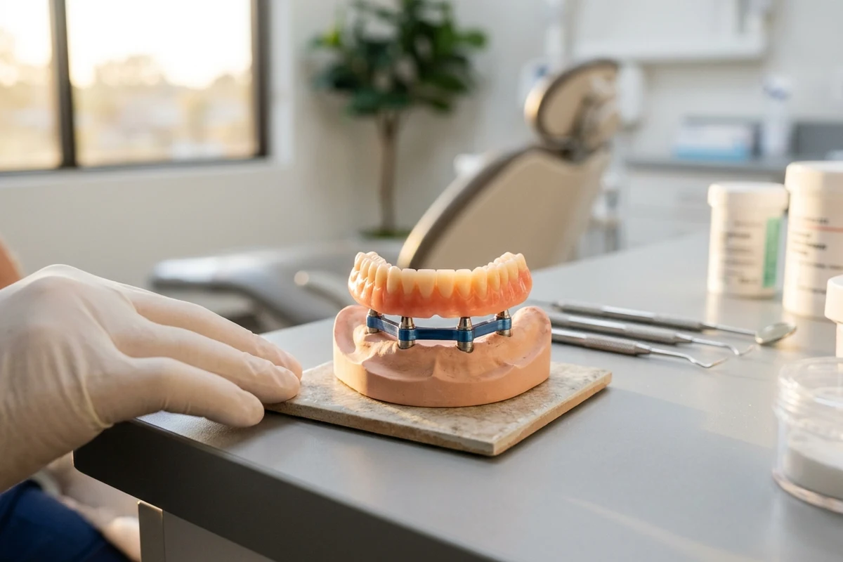 Restorative Care
Implant Supported Dentures Chino: Implant-Supported Dentures Fit and Maintenance
Implant-supported prostheses combine surgical anchors with specialized dental bases for enhanced chewing and retention. Understanding daily cleaning, component wear, and biological risks helps preserve your restoration and underlying bone for years to come.September 25, 2026 · 7 min read Emergency
Emergency Dentist Chino CA: Emergency Care for Dental Pain
When sudden pain, infection, or facial trauma strikes, consulting an emergency dentist Chino CA residents trust ensures rapid stabilization, accurate diagnostic imaging, and evidence-based interventions to relieve distress and preserve natural tooth structure.September 24, 2026 · 6 min read Preventive & General